
 EARS
 NOSE
 THROAT
 DIZZINESS
 TINNITUS
 SEARCH


What is the treatment for a cholesteatoma?
The treatment for a cholesteatoma is to remove it surgically. There are 3 main reasons to operate which are:
- To make the ear SAFE – to try and avoid the potentially serious complications
- To make the ear dry – a smelly discharge is the reason many patients come to the doctor
- To restore function – it maybe possible to repair any damaged hearing
What types of operations are there to remove a cholesteatoma?
There are many different ways to operate to remove a cholesteatoma and your surgeon will discuss his preferred technique with you. The approach chosen will be influenced by a number of factors.
There are many different ways to operate to remove a cholesteatoma and your surgeon will discuss his preferred technique with you. The approach chosen will be influenced by a number of factors but essentially there are two main types of technique:
1. OPEN techniques – also known as CANAL WALL DOWN techniques
and
2. CLOSED techniques – also known as CANAL WALL UP TECHNIQUES
The ‘canal wall’ referred to is the back wall of the ear canal. This area of bone separates the ear canal from the hollow mastoid bone.

CLOSED TECHNIQUES
Names for this type of operation:
- Canal Wall Up Mastoidectomy
- Combined Approach Tympanoplasty = a CAT
- Atticotomy with reconstruction
- Attico-antrostomy with reconstruction
My preferred option is the CLOSED TECHNIQUE.
In the CLOSED technique the mastoid bone is hollowed out to expose the cholesteatoma disease in the mastoid bone.
The ear drum is lifted up to expose the middle ear space and the disease in that area is removed.
The back wall of the ear canal is left in place separating the ear canal from the hollowed out mastoid bone. If areas of the ear canal need to be removed they are repaired with cartilage.
The ear drum is then rebuilt and placed back in its normal position. The patient is left with a normal ear canal.
The operation:

This is a right ear. The ear has been cut from behind and moved forward. The mastoid bone is exposed. The ear canal is the hole in front of the dotted line(arrow). The area to be drilled is shown by the dotted line.

The ear canal is widened a little to see the bony area of the ear canal which has been worn away. This is just above the ear drum(x) The mastoid bone is hollowed out and and the cholesteatoma disease can be seen.

The cholesteatoma ‘sac’ contains dead skin. It is like a sock with the opening above the ear canal. The dotted line represents the size of the sac. The sac has been opened and the skin inside can be seen.

More of the ‘sac’ has been removed – only the back wall is remaining. The ear canal remains intact.

The mastoid bone has been cleared of disease. The ear drum has been lifted up and the space behind the ear drum(arrow) has been cleared.
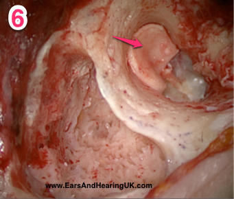
The deep part of the ear canal which was worn away by the disease has been rebuilt with cartilage(arrow)
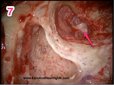
The ear drum has been rebuilt using a thin membrane of tissue called ‘fascia’ which comes from the covering of one of the muscles under the skin. The fascia continues up to cover the cartilage repair.
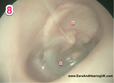
6 months after the operation. The new ear drum (A) and the repaired ear canal(B) can be seen. The patient now has a normal ear canal. They can get it wet and they can swim and it shouldn’t need regular cleaning. They can wear a hearing aid if necessary. Essentially they can forget about their ear and treat it as normal. If the hearing bones have been removed they can be replaced with a future operation.

The advantages of this technique:
- At that at the end of the operation the patient is left with a normal ear canal.
- By about two months after the surgery my patients can get the ear wet – so they can usually swim, shower and take part in water sports without the need for an ear plug.
- The ear drum is rebuilt and placed in its normal position recreating an air filled middle ear space. This allows attempts to restore the hearing at a future operation (occasionally this can be done at the first operation). Also after a year the ear drum is completely healed and its position can be gauged to allow an accurate assessment of the size of any prosthesis needed to reconstruct the hearing.
- Sometimes if the hearing chain has not been damaged it is possible to leave it in place and so preserve the hearing.
- Regular cleaning of the ear is not usually required.
- Ultimately the patient hopefully has a trouble free ear which they can completely forget about (apart from maybe an annual check by the surgeon)
The disadvantages are:
- That usually two operations are required the second operation being about a year after the first. This is to make sure that all the ‘skin’ was removed. Because the ear canal is till in place one can not see into the hollowed out mastoid bone by looking down the ear hole.If a few skin cells were left behind at the original operation they will very slowly regrow and form a small cyst. Because any regrowth is so slow we normally wait a year before the second operation to be sure that any regrowth will be big enough to see and remove. Sometimes an MRI scan can be performed instead of an operation to look for and regrowth of skin.
- The new ear drum may drop in and form a new ‘cholesteatoma’ which would need to be sorted out at the second operation.
OPEN TECHNIQUE
Names for this type of operation:
- Modified Radical Mastoidectomy
- Radical Mastoidectomy
- Canal Wall Down Mastoidectomy
In an OPEN technique the mastoid bone is hollowed out with a drill to expose the cholesteatoma, the ear drum is lifted up and the back wall of the ear canal, which separates the ear canal from the mastoid bone, is removed.
The hollowed out mastoid bone is joined to the ear canal. This creates a ‘mastoid cavity’.
The disease is removed, the ear drum is rebuilt and replaced inwards to close off the Eustachian tube.
The hollowed out mastoid bone is lined with a graft and the ear hole is enlarged. The patient is left with a ‘mastoid cavity’.

The operation:
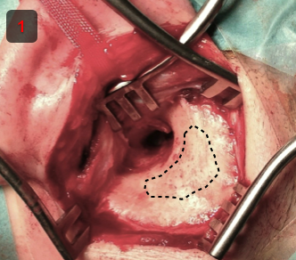
This is a left ear. The ear has been cut from behind and moved forward. The mastoid bone is exposed. The ear canal is the area in front of the dotted line. The area to be drilled is shown by the dotted line.
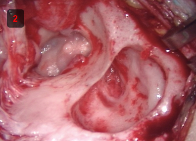
The mastoid bone is hollowed out and the disease is removed from the mastoid area
The bony wall between the ear canal and the mastoid cavity is removed.

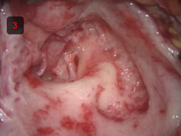
The hollowed out mastoid bone is now part of the ear canal. The ear drum is rebuilt to create a new middle ear space and separate the Eustachian tube from the mastoid cavity. The mastoid cavity grows a skin lining. The mastoid cavity can now be seen by looking into the ear hole.
The advantage of this technique is that it usually requires only one operation.
The disadvantages are:
- Takes longer to heal – often requiring regular cleaning of the cavity in the early post operative period which is often not well tolerated by children.
- The cavity may be prone to becoming infected.
- The cavity may build up wax and need lifelong regular visits to the doctor or nurse for dewaxing.
- Having the cavity cleaned with a suction machine can make the patient very dizzy because the balance system now form the back wall of the cavity.
- The cavity usually needs to be kept dry: meaning extra precautions when hair washng, showering and restricting water sports.
- If cold water or cold air gets in the cavity it can make the patient feel dizzy.
- It is more difficult to reconstruct the hearing (in my hands)
- It can be difficult to get a hearing aid to fit the enlarged ear hole; and closing off the cavity with a hearing aid can cause infections.
Having said all that some cavities are completely trouble free and self cleaning.
The above is my experience of open cavity techniques – other surgeons may have a different experience and some employ techniques to make the cavity smaller and try and overcome these problems.
What does the operation involve?
The operation is performed under a general anaesthetic – so you will be fast asleep and not be aware of what is going on.
Sometimes a very small amount of hair needs to be shaved from behind the ear.
I make a cut in the crease behind the ear which will be closed with absorbable stitches at the end of the operation.
The operation could take anything between 2 – 4 hours depending on the extent of the disease. There are lots of delicate structures all within a few millimetres of each other so great care is taken to avoid any injury to these structures.
At the end of the operation the ear canal or the mastoid cavity is packed with an antiseptic dressing. This will stay in place for about 3 weeks and is removed in the outpatients clinic.
You will usually wake up with a large bandage around your head which is holding a pressure dressing over the ear. The bandage will be removed after a few hours or the following morning.
Patients used to always stay overnight and go home the next dat but we are performing more operations as day cases so you go home on the same day as the operation. This will depend on the timing of the surgery, the duration of the surgery, how you feel afterwards, your personal preference and your home circumstances.
You can not drive for 48 hours after any general anaesthetic operation (your car insurance will not be valid).
The area may feel uncomfortable afterwards but is not usually particularly painful. The discomfort is usually controlled with simple painkillers.
I will see you in the outpatients department 3 – 4 weeks after the surgery and remove the ear canal dressing. You will then need some further visits to make sure that everything is healing as planned.
It is important to keep water out of the ear canal until the area if fully healed. Most of my patients who have had a closed technique procedure will be able to get the ear wet after about 2 months. An open technique procedure may take longer to heal and the ear may always need to be kept dry.
I suggest that you do not fly for about 2 months after the operation.
What are the risks of the operation?
The ear contains many different important structures in a very confined space.
Any of these structures could be damaged by the untreated cholesteatoma disease or the operation.
These complications of the untreated disease are uncommon but potentially very serious.
All of the structures in the ear are also at risk during the operation. Below is an information sheet that I give to all my patients to explain the risks of operations for cholesteatoma.

Is there an alternative to surgery?
The alternative to surgery is to continue with regular cleaning and observation in the outpatient clinic, which I would not normally recommend.
Surgery is not without risk and the alternative to surgery is to continue with regular cleaning and observation in the outpatient clinic. In the case of cholesteatoma this is not an alternative that I would normally recommend.
Looking down the ear canal in the outpatient clinic does not allow us to see the extent of the disease or completely remove it with the suction equipment. If the cholesteatoma disease is left untreated it could damage any of the structures discussed above. Chronic infection could spread into the mastoid bone causing a mastoiditis or deeper into the head causing meningitis of brain abscess. These are rare but serious complications.
IT MUST BE STRESSED THAT THESE ARE VERY GENERAL PRINCIPLES.
EVERYONE IS DIFFERENT AND THERE ARE MANY VARIATIONS ON WHAT IS DESCRIBED HERE.
EACH PATIENT SHOULD BE TREATED AS AN INDIVIDUAL.
THE APPROPRIATE TECHNIQUE OR COMBINATION OF TECHNIQUES SHOULD BE CHOSEN DEPENDING ON A PATIENTS INDIVIDUAL CIRCUMSTANCES AND THE WIDE VARIETY OF FACTORS WHICH INFLUENCE THAT PATIENTS TREATMENT.
IF YOU ARE NOT ONE OF MY PATIENTS I AM SURE YOUR SURGEON WILL DISCUSS HIS PRACTICE WITH YOU IN DETAIL.
PLEASE DO NOT BASE TREATMENT DECISIONS ON THE CONTENTS OF THIS WEBSITE. TALK TO YOUR DOCTOR.
